(last modified October, 2006)
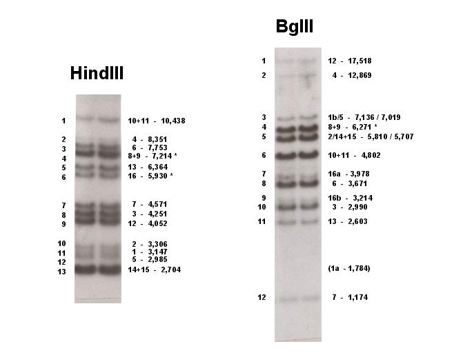
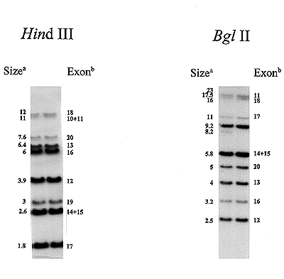
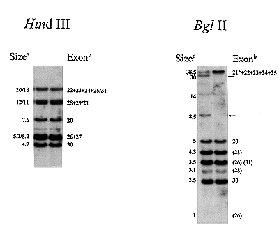
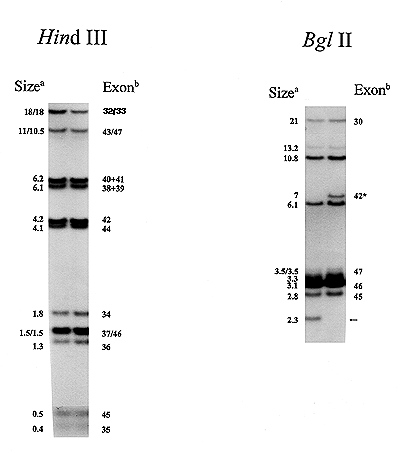
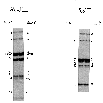
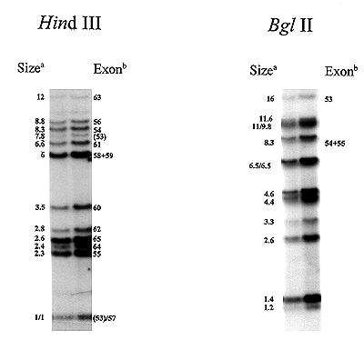
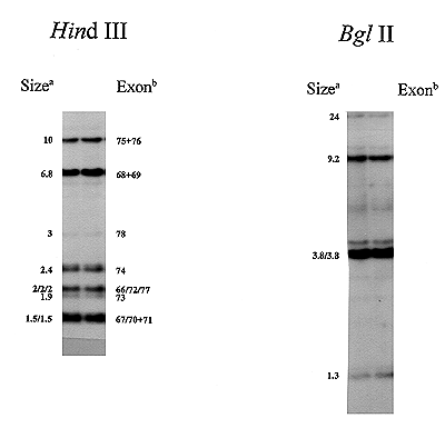
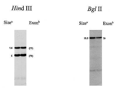
These pages show images of Southern blots of a human genomic DNA digested with HindIII or BglII after hybridisation with a series of DMD cDNA's, together spanning the entire DMD coding region. Images were kindly provided by Sander Kneppers and ... (Human and Clinical Genetics, Leiden University Medical Center, LEIDEN, Nederland).
For origin and content of the cDNA-probes see DMD cDNA probes.
Southern blot of a human genomic DNA digested with HindIII (left) or BglII (right). Fragment numbers are+ indicated on the left of each panel, the exon number and the size of the fragment (in bp) on the right. 1+2 = two exons located on one fragment, 1a / 1b = the two halves of an exon digested by the restriction enzyme used, (1a - 1,174) = a fragment hybridising very weekly (might not be visible), * = a polymorphism has been reported for this band (see The DMD cDNA in relation to the gene).
| Top of page | LMDp home page
|
| Remarks / information | Copyright©,
liability |