(Judith van Deutekom/Anneke Janson; last modified on March 25, 2000)
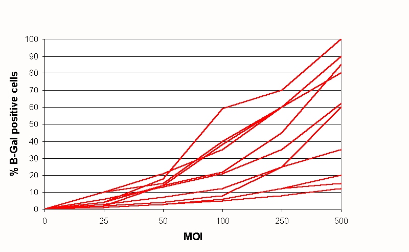
Figure 3
Adenoviral infection of different chorion villi cell cultures. Ten different
chorion villi cell cultures were infected with an adenoviral vector carrying the LacZ gene
(Ad5-LacZ), at increasing MOIs. The results show the high variability in adenoviral
transduction efficiencies in the individual cell cultures, determined as the percentage of
beta-galactosidase positive cells.
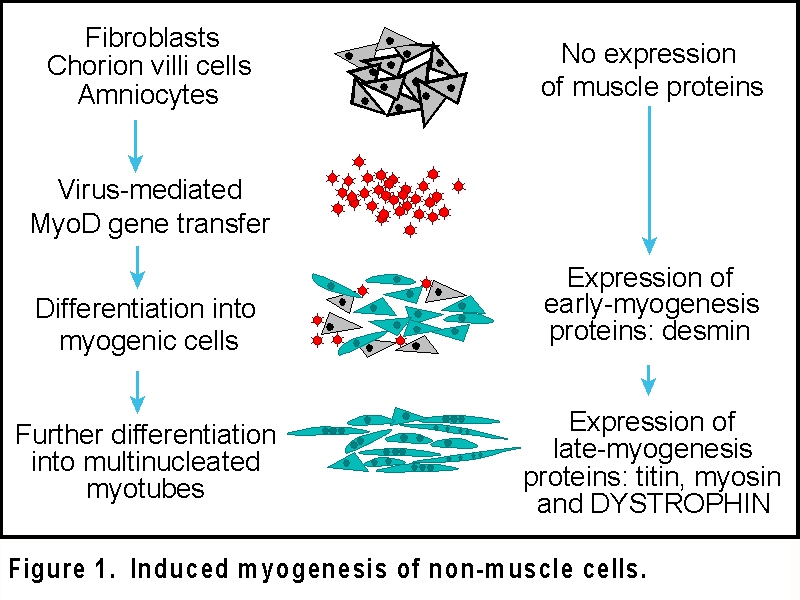
Note the appearance of multi-nucleated cells, indicating successful differentiation towards muscle-like cells.
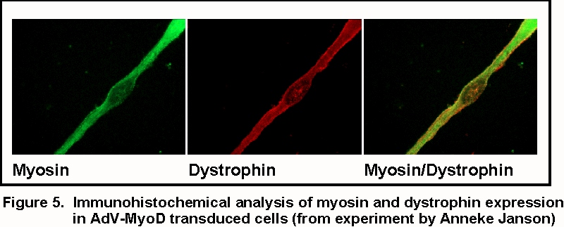
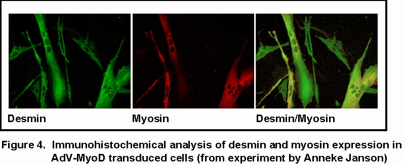
Note that the myosin staining is througout the cell, while dystrophin staining is more in the cell membrane.
Dr. Judith C.T. van Deutekom
Department of Human and Clinical Genetics,
Leiden University Medical Center
E-mail: deutekom@lumc.nl
/ Tel: +31 - 71 - 527 6080
Dr. Johan T. den Dunnen
Department of Human and Clinical Genetics,
Leiden University Medical Center
E-mail: ddunnen@lumc.nl
/ Tel: +31 - 71 - 527 6105
| Top of page | MyoD
homepage | LMDp homepage |
| Diagnostic techniques | Remarks / information | Disclaimer |